Craniomaxillofacial
Maxillofacial surgical solutions
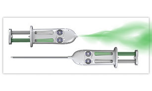
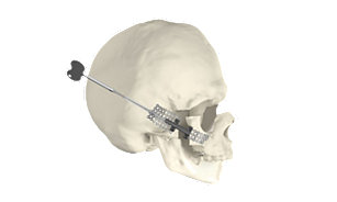
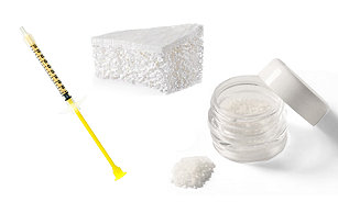
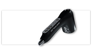
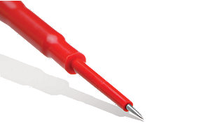
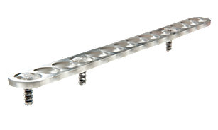
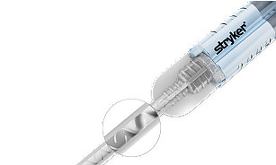
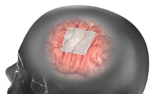
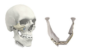
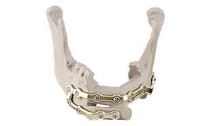
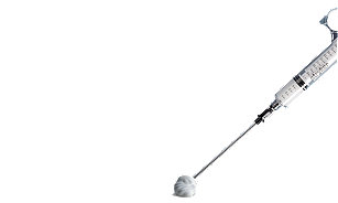
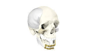
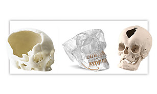
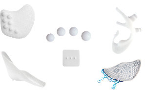
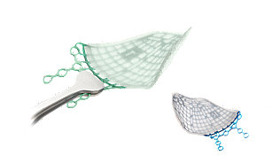
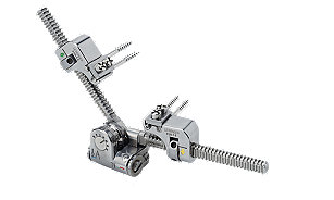
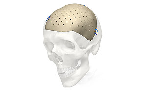
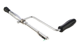
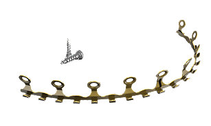
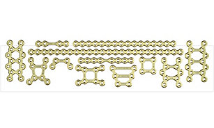
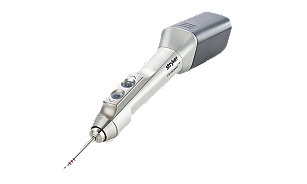
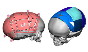
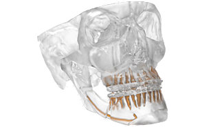
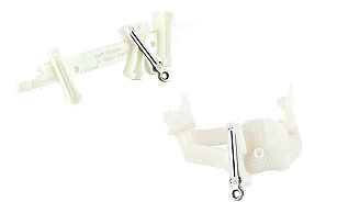
Your patients deserve best in class products delivered by people who care. Our comprehensive portfolio features rigid bone fixation systems, biomaterials, porous polyethylene implants, microdissection needles and patient-specific implants to address a wide variety of surgical specialties and procedures.

CMF-WC-28_Rev. None_14666